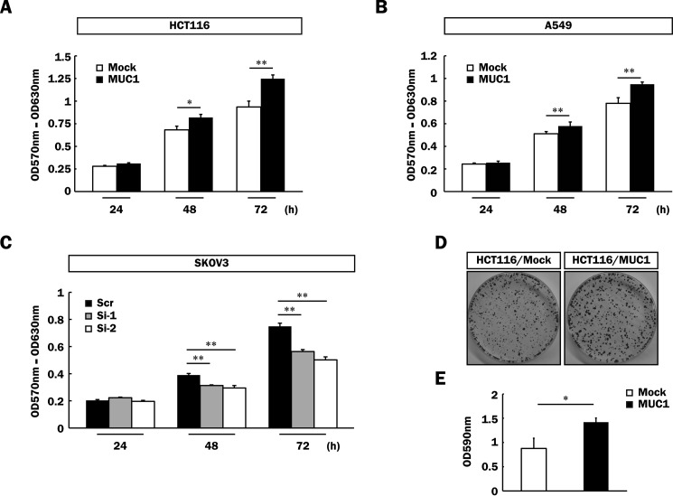
FIGURE 8.
Enhancement of cell proliferation of various MUC1-expressing cells. A–C, HCT116/Mock and HCT116/MUC1 (5 × 10³ cells) (A), A549/Mock and A549/MUC1 (2 × 10³ cells) (B), and SKOV3/Scr, SKOV3/Si-1, and SKOV3/Si-2 cells (2 × 10³ cells) (C) were plated and cultured for 24, 48, and 72 h. The level of cell proliferation was assessed by MTT assays (means ± S.D., n = 3). *, p < 0.05; **, p < 0.01. D, HCT116/Mock and HCT116/MUC1 cells (5 × 10³ cells) were plated and cultured for 2 weeks. The cells thereafter were fixed and stained with crystal violet as described under “Experimental Procedures.” E, the dye was extracted from the cells as described under “Experimental Procedures,” and its level was determined by measuring the absorbance at 590 nm (means ± S.D., n = 3). *, p < 0.05.

