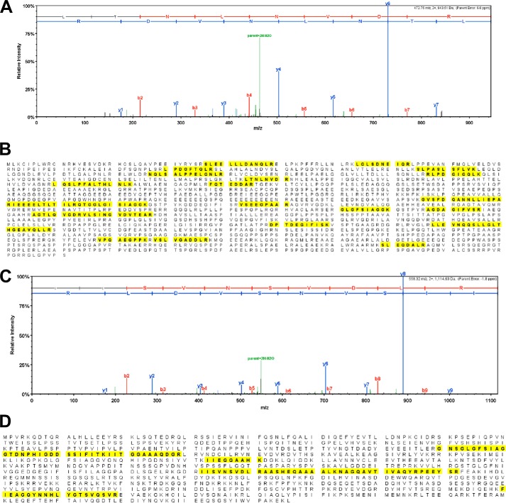
FIGURE 3.
MS/MS analysis to show hScrib and DLG-1 binding to STax1. A, representative MS/MS data for one of the tryptic peptides (LTNLNVDR) from hScrib is shown. B, identified peptide sequences in hScrib are indicated in bold and highlighted in yellow. The amino acid sequence coverage was 15% (249/1631 amino acids) with 22 unique peptides and 47 total spectra being identified in fractions with STax1. No peptides from hScrib were detected in fractions with STax2 or STax1ΔPBM. C, representative MS/MS data for one of the tryptic peptides (IISVNSVDLR) from DLG-1 is shown. D, identified peptide sequences in DLG-1 are indicated in bold and highlighted in yellow. The amino acid sequence coverage was 12% (110/926 aa) with 7 unique peptides and 13 total spectra being identified in fractions with STax1. No peptides from DLG-1 were detected in fractions with STax2 or STax1ΔPBM.

