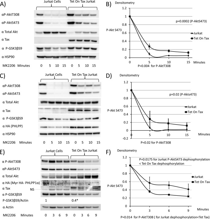
FIGURE 8.
Expression of membrane-associated PHLPP overcomes the effects of Tax1 on rates of Akt dephosphorylation. A and B, Jurkat cells and Tet-On Tax1 Jurkat cells maintained in doxycycline for 48 h were treated with the Akt inhibitor MK2206, and samples were taken at serial time points to assess levels of P-AktThr-308(P-AktT308), P-AktSer-473 (P-AktS473), total Akt, Tax1, P-GSK3βSer-9 (P-GSK3βS9), and HSP90. The levels of P-AktSer-473 at each time point were normalized to values obtained at 0 min of MK2206 treatment. C and D, doxycycline-treated Jurkat cells and Tet on Tax1 Jurkat cells were transiently transfected with a PHLPP expression vector for 6½ h and then treated with MK2206, and samples were taken at serial time points to compare Akt dephosphorylation rates, as described above. E and F, doxycycline-treated Jurkat cells and Tet-On Tax1 Jurkat cells were transfected with a myristoylated PHLPP expression plasmid for 6½ h and then treated with MK2206, and samples were taken at serial time points to compare Akt dephosphorylation rates, as described above. Dephosphorylation curves were generated from two independent experiments. The baseline P-AktSer-473 level is normalized to 1 for dephosphorylation curves.

