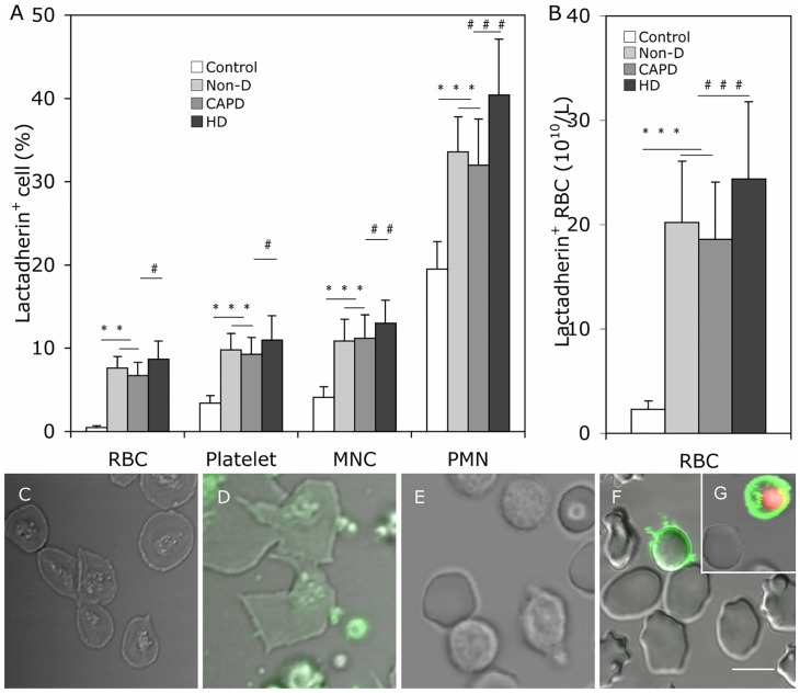
Fig 2. PS exposure on the plasma membrane of blood cells.
Comparison of PS exposure on blood cells among Non-D patients, CAPD patients, HD patients and healthy subjects. Cells were incubated with Alexa Fluro 488 -lactadherin separately in the dark for 10 min at room temperature before evaluation by flow cytometry. (A) We measured the percent of RBCs, platelets, PMNs, MNCs that bound lactadherin from healthy subjects (n = 20), Non-D (n = 25), CAPD (n = 18) and HD patients (n = 23). (B) Lactadherin -binding RBC number per liter of plasma according their count in each person. Data are expressed as mean ± SD (***P < 0.001, #P < 0.05, ##P < 0.01, ###P < 0.001). PS exposure on the plasma membrane of blood cells was observed by confocal microscopy with LSM 510 3.2 SP2 software. Platelets, RBCs and WBCs of healthy subjects and uremic patients were incubated with Alexa Fluro 488 -lactadherin and PI in the dark 10 min at room temperature. Cells were then washed very gently to remove unbound dye. Cell membrane displayed green fluorescence when labeled with lactadherin and nucleus displayed red fluorescence. Lactadherin staining (green) is observed on platelets membrane and MPs (D) and RBC (F)/WBC (G) in uremic patient but no staining in healthy subjects (C, E). The inset bar equals 5 μm. PS: phosphatidylserine; PMN: polymorphomuclear cell; MNC: mononuclear cell; Non-D: Non-dialysis; CAPD: continuous ambulatory peritoneal dialysis; HD: haemodialysis.

