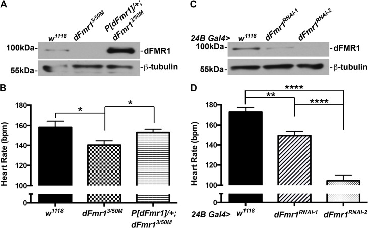
Fig 1. dFmr1 regulates heart rate.
(A) Western blot analysis showing the complete knockout of dFmr1 (dFmr1 3/50M) compared to wild type (w 1118) and genomic rescue (P[dFmr1]/+; dFmr1 3/50M). (B) Loss of dFmr1 (dFmr1 3/50M, N = 16) results in a statistically significant decrease in heart rate when compared to wild type (w 1118, N = 10). This is rescued by expression of a wild type copy of dFmr1 in its genomic context (P[dFmr1]/+; dFmr1 3/50M, N = 10). (C) Western blot showing that two independent RNAi lines (24B Gal4> dFmr1 RNAi-2, dFmr1 RNAi-2) result in almost complete striated muscle-specific knockdown of dFMR1 (i.e., ~85–100%) compared to control (24B Gal4> w 1118). (D) Pan-muscle (24B Gal4) RNAi knockdown of dFmr1 (24B Gal4> dFmr1 RNAi-1, N = 12 and dFmr1 RNAi-2, N = 6) leads to a significant decrease in heart rate compared to control (24B Gal4> w 1118, N = 17). Student’s T-test was used to calculate statistical significance: p values, * <0.05, ** <0.01, **** <0.0001.

