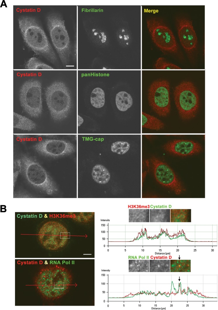
FIGURE 3.
Intranuclear location of cystatin D in SW480-ADH cells. A, double immunolabeling for cystatin D in combination with fibrillarin (nucleolus marker, upper panel), pan-histone (chromatin marker, middle panels), and TMG-cap (nuclear speckles and Cajal bodies marker, lower panels). Scale bar, 5 μm. B, upper panels, double immunolabeling for cystatin D and histone H3K36me3, epigenetic marker of transcriptionally active chromatin. The enlarged insets show a representative example of a nuclear domain with co-localization foci of both immunostaining signals. The graph shows a line profile analysis of signal intensities (arbitrary units) from histone H3K36me3 (red) and cystatin D (green) across a line. Lower panels, double immunostaining analysis of cystatin D and active RNA pol II expression. The enlarged inset shows a magnification of a representative nuclear domain illustrating the concentration of both fluorescent signals in microfoci. The graph shows an intensity profile of cystatin D (red) and RNA pol II (green) across a line revealing the co-localization of active RNA pol II and cystatin D in a nuclear microfocus (arrow), whereas the latter was absent from other RNA pol II-positive microfoci. Scale bars, 2.5 μm.

