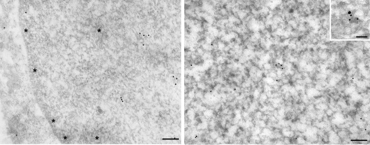
FIGURE 4.
Immunogold electron microscopy location of cystatin D in the nuclei of SW480-ADH cells. Gold particles appear distributed in euchromatin domains, whereas condensed chromatin areas (asterisks) are devoid of labeling. Inset, double immunogold labeling revealed the colocalization of RNA pol II (15-nm particle) and cystatin D (10-nm particles) in a nuclear microfocus within euchromatin. Scale bars, 100 nm; inset, 50 nm.

