FIGURE 4.

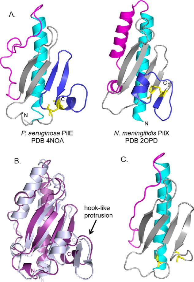
Comparison of PilEΔ1–28 with N. meningitidis PilXNm. A, side-by-side comparison of PilEΔ1–28 and PilXNm,Δ1–28 with the N-terminal α-helices colored in cyan, αβ-loops in magenta, β-sheets in gray, and D-regions in blue with the cysteines represented as sticks in yellow. B, structural alignment of PilEΔ1–28 (purple) and PilXNm,Δ1–28 (light blue). 104 residues are aligned with an RMSD of 4.3 Å. C, Phyre2-generated model of PilVNm based on PilE. Structural illustrations and alignments were generated with PyMOL (version 1.3, Schrödinger, LLC.).
