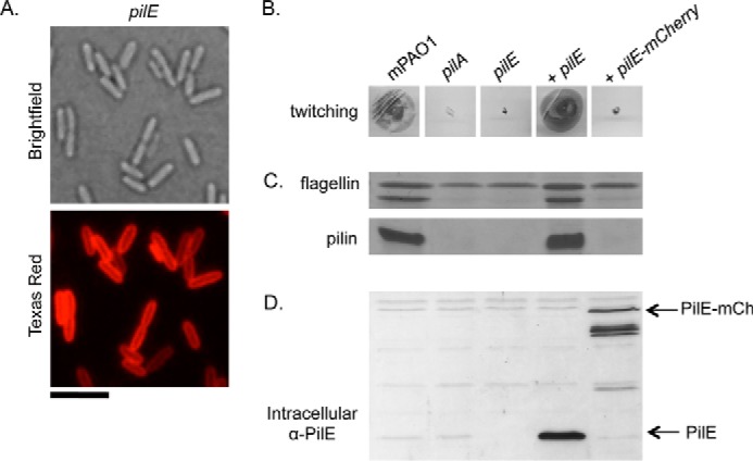
FIGURE 5.

Complementation of pilE with PilE mCherry. mCherry was fused to the C-terminal end of PilE, and the level of complementation of a pilE mutant was assessed. A, fluorescence microscopy analysis of PilE mCherry localization. Scale bar represents 5 μm. B, twitching motility was tested by stab-inoculating to the bottom of an LB 1% agar plate and staining with 1% crystal violet after a 24-h incubation at 37 °C. C, pili were sheared from the surface of cells of interest and separated on a 15% SDS-PAGE gel. The flagellin band is used as a loading control. D, intracellular levels of PilE were probed by Western blot analysis with a α-PilE peptide antibody (1:1000 dilution). Arrows indicate the bands of interest.
