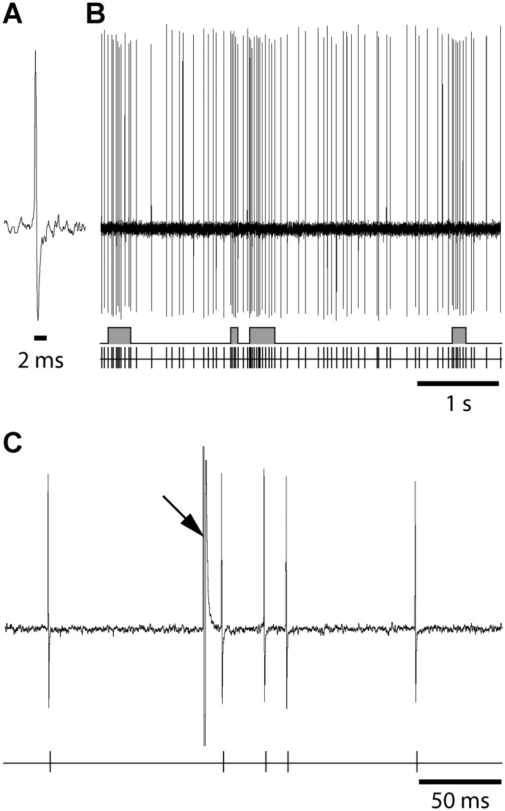
Fig 2. Raw recording traces from a SNr cell illustrating the short spike duration (A), its spontaneous discharge (B) and its response to one single orofacial motor cortex stimulation (C).
(A) Magnified view of recording traces from a SNr neuron illustrating that its spikes are narrow (< 2 ms). (B) The spontaneous activity is displayed following injection of haloperidol; the discharge of the cell is represented as the raw recording trace (top), the result of the Poisson Surprise spike analysis (S ≥ 2) indicating bursts (middle) and the corresponding sequence of spikes (bottom). (C) Response evoked by a single cortical stimulation illustrated as a raw trace (top) and the corresponding sequence of spikes (bottom). Arrow indicates the artifact stimulation.

