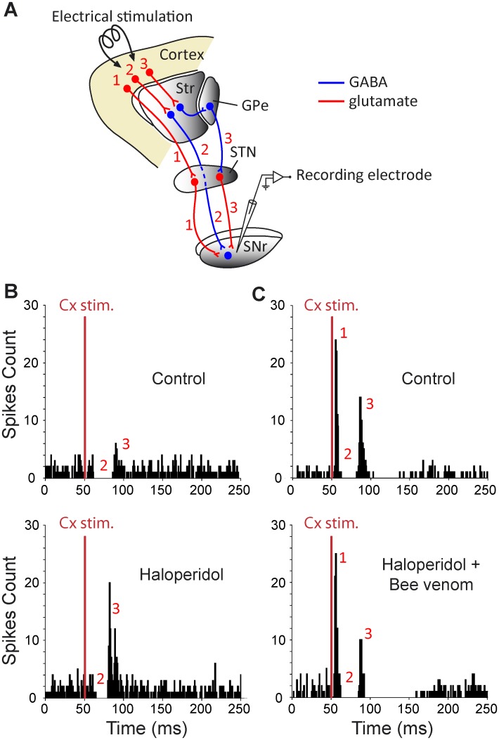
Fig 4. Experimental design (A) and effect of systemic injection of haloperidol alone (B) or followed by bee venom injection (C) on cortically-evoked responses on SNr neurons.
(A) Schematic representation illustrating the three main basal ganglia pathways activated by cortical stimulation and connecting the cerebral cortex to the SNr. It includes the hyperdirect cortico-subthalamo-nigral pathway (#1) and the direct (#2) and the indirect (#3) striato-nigral pathways. Str, striatum; GPe, external globus pallidus; STN, subthalamic nucleus. (B, top) In this SNr neuron, orofacial sensori-motor cortex stimulation in control conditions evoked a complex response composed of an inhibition followed by a late excitation. (B, bottom) 60 minutes after haloperidol injection (1 mg/kg), the late excitation of the cortically-evoked response was markedly increased. (C, top) Classical triphasic excitatory-inhibitory-excitatory sequence evoked by stimulation of the orofacial sensori-motor cortex in control conditions in another SNr cell. In this cell, the increase of the late excitatory component was prevented by injecting BV 30 minutes after haloperidol as shown by comparing the cortical responses elicited 60 minutes after haloperidol (C, bottom) to the control response of the same SNr cell (C, top). Red numbers in B-C indicate which pathway in A is responsible for each component (excitation-inhibition-excitation) of the cortically-evoked responses. The same number of cortical stimulations (red bar, n = 50) was applied in B-C.

