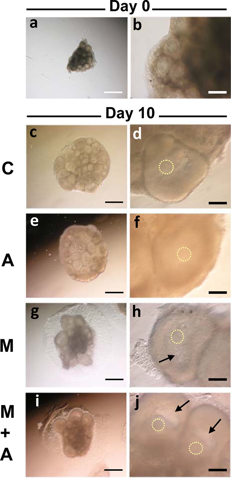
Fig 3. Morphology of ovarian tissues cultured under 4 different conditions.

(a, b) A whole murine ovary at 14 days old was cut into 8 pieces before culture (a). The most advanced follicles in the tissue was at the preantral stage (b). (c-j) Ovarian tissues after 10 days of culture in the C (c, d), A (e, f), M (g, h) and M+A (i, j) conditions. The antral cavity formation occurred only in the follicles under the 3-D Culture system (M, M+A) (arrows). The circles of dots represent the localization of oocytes. Regular bar = 500 μm. Bold bar = 100 μm.
