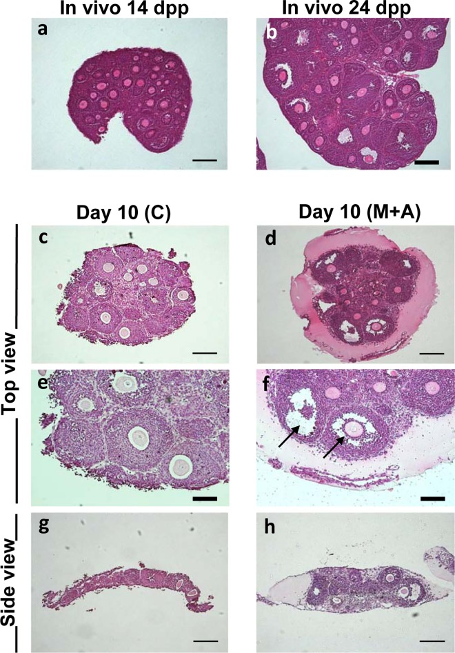
Fig 4. Histological analysis of ovarian tissues under Membrane or 3-D culture system.
(a, b) Paraffin sections of a whole murine ovary at 14 dpp (a) and 24 dpp (b) with a HE staining. (c, e, g) The ovarian tissues cultured in the Membrane culture system (C condition) for 10 days. (d, f, h) The ovarian tissues cultured in the 3-D culture system (M+A condition) for 10 days. Follicle growth was detected in both culture systems. Importantly, the antral cavity formation (arrows) occurred only in the 3-D culture system (M, M+A) regardless of the presence or absence of activin A treatment. Regular bar = 200 μm. Bold bar = 100 μm.

