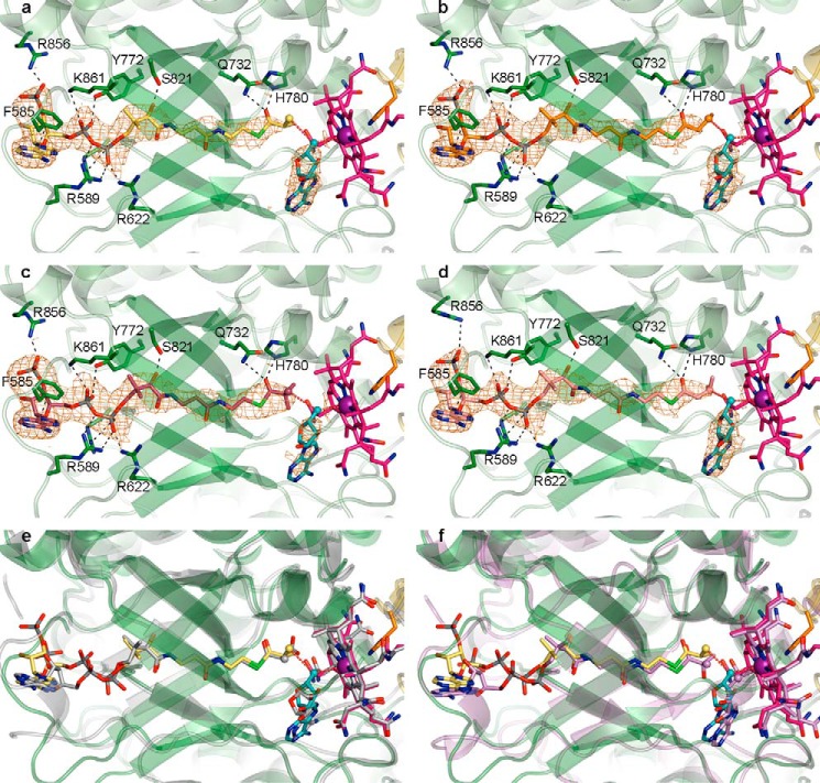
FIGURE 4.
Overall mode of substrate binding in IcmF and comparison with MCM and HCM1. a, IcmF bound to Cbl (pink carbons; cobalt, purple), 5′-dAdo (cyan carbons), and isobutyryl-CoA (yellow carbons). 2Fo − Fc omit electron density (orange mesh) contoured at 1.0 σ is shown around the substrates and 5′-dAdo. IcmF is shown with the chain A TIM barrel substrate-binding domain in green and the Cbl-binding domain in yellow. The Cbl-coordinating His-39 as well as selected residues that bind substrate are shown as sticks, with hydrogen bonds, ionic interactions, and π-π interactions shown as black dashed lines. The 5′-dAdo C5′ and the locations of hydrogen abstraction on the substrates are shown as spheres, and red dashed lines connect the C5′ to the Cbl cobalt and the substrate carbons. b, IcmF bound to n-butyryl-CoA (orange carbons) displayed as in a. c, IcmF bound to pivalyl-CoA (maroon carbons) displayed as in a. d, IcmF bound to isovaleryl-CoA (light pink carbons) displayed as in a. e, comparison of isobutyryl-CoA-bound IcmF (green) and methylmalonyl-CoA-bound MCM (gray, PDB code 4REQ), revealing nearly identical structures and substrate binding modes. IcmF shown as in a. MCM-bound methylmalonyl-CoA, Cbl, and 5′-dAdo are shown with gray carbons. f, comparison of isobutyryl-CoA-bound IcmF (green) and hydroxyisobutyryl-CoA-bound HCM1 (lilac, PDB code 4R3U), revealing similar structures and substrate binding modes. IcmF shown as in a. HCM1-bound hydroxyisobutyryl-CoA, Cbl, and 5′-dAdo are shown with lilac carbons.

