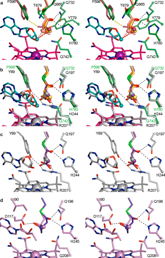
FIGURE 7.
IcmF active site and comparison with MCM and HCM1 in wall-eyed stereo view. a, overlay of four substrate-bound IcmF structures, revealing similar substrate orientation. Shown are isobutyryl-CoA (yellow carbons), n-butyryl-CoA (orange carbons), pivalyl-CoA (maroon carbons), and isovaleryl-CoA (light pink carbons). The locations of hydrogen abstraction are shown as spheres, located within 3.6 Å of the 5′-dAdo (C3′-endo conformation, cyan carbons) C5′ atom (cyan sphere), as indicated by the dashed red lines. Residues in the substrate-binding site are shown with dark green carbons. The third methyl group of pivalyl-CoA clashes with Phe-598 (yellow dashed line), leading to a small rotation of the side chain (maroon carbons) in this structure. Hydrogen bonds from Gln-732 and His-780 to the thioester carbonyl are indicated as dashed black lines. Cbl is shown with carbons in pink and cobalt in purple. b, overlay of substrate-bound IcmF and MCM (26) (PDB code 4REQ). Isobutyryl-CoA, n-butyryl-CoA, IcmF-bound 5′-dAdo, IcmF-bound Cbl, and IcmF residues are shown as in a. The locations of hydrogen atom abstraction in both methylmalonyl-CoA (gray carbons) and succinyl-CoA (purple carbons) overlay with those of IcmF substrates (spheres). MCM-bound 5′-dAdo, MCM-bound Cbl, and MCM substrate-binding residues are shown with gray carbons. Gln-197 and His-244 are conserved in MCM and IcmF, whereas IcmF Phe-598 is replaced by MCM Tyr-89 and IcmF Gln-742 is replaced by Arg-209, putatively accounting for the switch in substrate binding specificity. Hydrogen bonds and ionic interactions are shown as dashed black lines. c, active site of substrate-bound MCM, shown as in b, highlighting interactions to substrate carboxylate groups. Only interactions to methylmalonyl-CoA are shown for clarity. d, active site of substrate-bound HCM1 (PDB code 4R3U) (29), highlighting interactions to substrate hydroxyl groups. Hydroxyisobutyryl-CoA is shown in lilac and (S)-3-hydroxybutyryl-CoA in blue. Only interactions to hydroxyisobutyryl-CoA are shown for clarity. Locations of hydrogen atom abstraction are shown as spheres.

