
FIGURE 1.
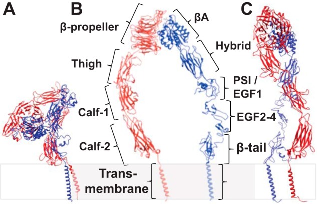
Schematic structure of integrin αIIbβ3. In the inhibited state (A), the integrin adopts a bent-over conformation, whereas when integrin is in the active state (B and C), it adopts an up-right, extended conformation. When the leg densities are in the same plane, they appear to be separate (B). However, if the view is from the side, the leg densities could appear crossed-over (C) even though they come from the same structure with widely separated legs when viewed from a different direction. Because there is no high resolution structure of full-length integrin αIIbβ3, details of the domain interaction and placement are imprecise. Panel A was generated using PDB structures 1JV2 (10) and 2K9J (51); panels B and C were generated using PDB structures 2VDM (49), 2P28 (52), 2K1A (53), and 2MRZ (53).