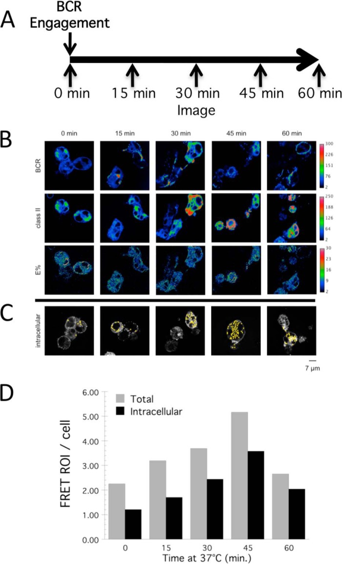
FIGURE 4.
Association of BCR and MHC class II molecules within intracellular compartments. A, time line of the experimental protocol. B, representative images of anti-BCR-pulsed FRET cells from each time point. Rows from top to bottom: CD79a-YFP (BCR), I-Ak-CFP (class II), corrected FRET between CD79a-YFP, and class II-CFP (E%). C, representative images with identified intracellular FRET events. D, quantitation of the number of FRET events/cell at each time point across all samples analyzed. Both total FRET events/cell (Total) and morphologically identifiable intracellular FRET events/cell (Intracellular) are reported.

