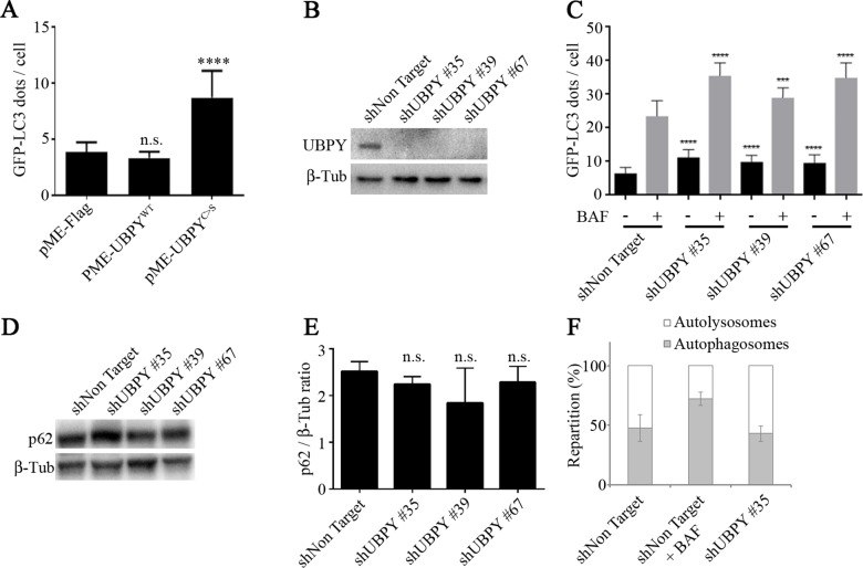
Fig 6. UBPY silencing in HeLa cells activates autophagy.
(A) The number of GFP-LC3 dots per cell was quantified in HeLa cells stably expressing the autophagy reporter GFP-LC3; cells were transfected with a control plasmid (pME-Flag) or plasmids expressing either the wild-type human UBPY protein (pME-UBPYWT) or its catalytically inactive mutant (pME-UBPYC>S). Bars denote mean ± s.d. Statistical significance was determined using t-test: ****p<0.0001 (B) The expression of UBPY was monitored by Western blot in GFP-LC3 HeLa cells stably transfected with a control shRNA or three different shRNAs targeting UBPY. (C) The number of GFP-LC3 dots per cell was quantified in GFP-LC3 HeLa cells stably transfected with a control shRNA or three different shRNAs targeting UBPY in absence (black bars) or in presence of bafilomycin A1 (BAF, gray bars). Bars denote mean ± s.d. Statistical significance was determined using t-test: ****p<0.0001; ***p<0.005 (D) The expression of the autophagy target protein p62 was monitored by Western blot in GFP-LC3 HeLa cells stably transfected with a control shRNA or three different shRNAs targeting UBPY. (E) Quantification of p62 levels in GFP-LC3 HeLa cells stably transfected with a control shRNA or three different shRNAs targeting UBPY from three independent Western blots. (F) The repartition of autolysosmes and autophagosomes was determined in mRFP-GFP-LC3 HeLa cells stably expressing either the control shRNA or the shUBPY #35 shRNA, in comparison with control transfected cells treated with bafilomycin A1.

