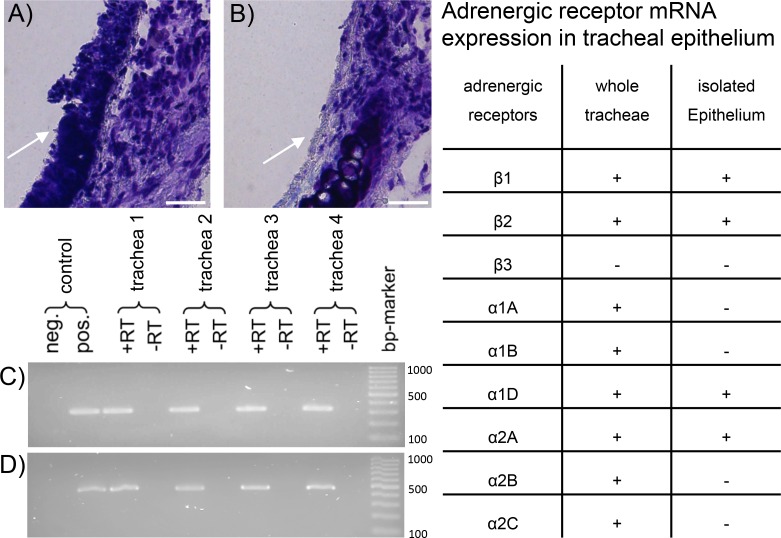
Fig 1. Adrenergic receptor mRNA expression in tracheal epithelium.
A: Transversal section of untreated murine trachea displays complete and intact respiratory epithelium (arrow) as used for PTV experiments and for mRNA analysis from entire organs. B: Tracheal wall section after isolation of epithelium by brush removal shows the specificity of epithelial cell isolation for mRNA extraction. Thus, the arrow indicates a section of the trachea where the epithelium remained. All other tracheal wall structures were left intact. C & D: In isolated epithelium harvested from four different tracheae (1–4) mRNA of the adrenergic receptors β1(C), β2 (D) were found (α1D and α2A are not shown). epithelium: → scale bars: 25 μm. Table: Distribution of adrenergic receptors in the whole tracheae and in the isolated epithelium. +: mRNA expressed; -: mRNA not detectable.

