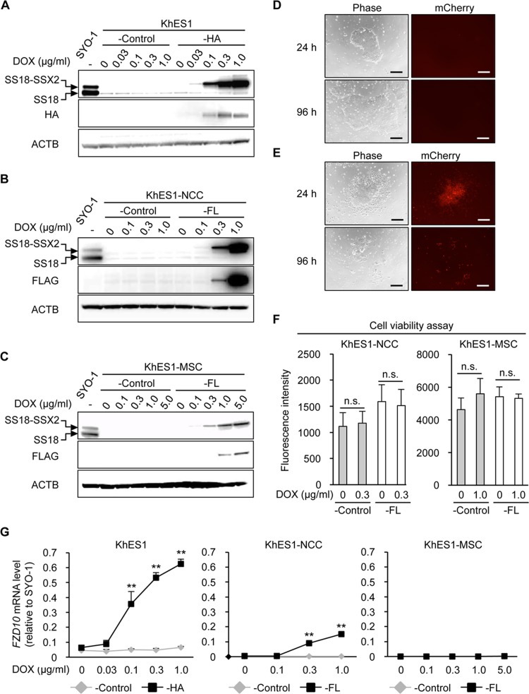
Fig 2. Induction of SS18-SSX2 in hESCs, hNCCs, and hNCC-derived MSCs.
A-C) DOX dose-dependently induced the SS18-SSX2 protein in KhES1-HA (A), KhES1-NCC-FL (B), and KhES1-MSC-FL (C) cells. Cells with Stuffer (-Control) and SS18-SSX2 were treated with the indicated concentrations of DOX for 24 h, and the expression of SS18-SSX2 was analyzed by Western blotting. The SS18-SSX2 and SS18 proteins were detected using an anti-SS18 antibody (top panel), and the 3xHA-SS18-SSX2 or FLAG-SS18-SSX2 protein was detected by an anti-HA or anti-FLAG antibody (middle panel). D and E) Morphology (left panels) and expression of mCherry (right panels) in KhES1-HA cells treated with 0 (D) or 0.3 (E) μg/ml of DOX for 24 and 96 h. Scale bar, 200 μm. F) Effects of SS18-SSX2 on the cell viability of KhES1-NCC-FL and KhES1-MSC-FL cells. Cells with Stuffer (-Control) or SS18-SSX2 were treated with the indicated concentrations of DOX for 48 h, and cell viability was measured using the AlamarBlue assay. n.s. means not significant. Error bars reflect SD in 4 experiments. G) Induction of FZD10 expression by SS18-SSX2 in KhES1-HA, KhES1-NCC-FL, and KhES1-MSC-FL cells. Cells with Stuffer (-Control) or SS18-SSX2 were treated with the indicated concentrations of DOX for 24 h, and the expression of FZD10 was analyzed by RT-qPCR. Expression levels were normalized to those of human ACTB and calculated as fold changes relative to SYO-1. Error bars reflect SD in 3 experiments. **, p<0.01 by the t-test.

