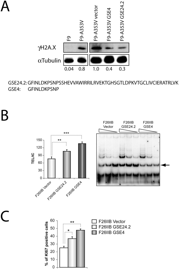
Fig 1. DNA-damage protective effect, telomerase activation and cell proliferation induction of one small peptide, GSE4, derived from GSE24.2.
Panel A. One small peptide derived from GSE24.2, GSE4, and GSE24.2 were expressed in F9_A353V cells that were transfected with the pRRL-CMV-IRES-EGFP vector, either empty (Vector) or expressing GSE24.2 or GSE4. Twenty four hours later cells were lysed and the presence of γH2AX and α-tubulin (loading control) analyzed by western blot. Un-transfected F9 and F9-A353V cells were used as controls. The values at the bottom of the panel indicate the estimated ratio between γH2AX and α-tubulin expression levels referred to those found in cells transfected with the empty vector (F9-A353V vector). The amino acid sequences of GSE24.2 and GSE4 are indicated at the lower part of the panel. Panel B. The telomerase activity of F26IIB cells transfected with the pRRL-CMV-IRES-GFP vector empty (vector), expressing GSE24.2 (GSE24.2) or GSE4 (GSE4) was determined using the Telomeric Repeat Amplification Protocol (TRAP) assay. The amplification products obtained using three decreasing amounts of cell extracts for each cell line are shown in the right panel. Quantification of the amplification products, normalized to the internal control provided in the assay (indicated by an arrow at the right panel) is shown in the left panel. Panel C. Expression of Ki67 was determined by immunocytochemistry in F26IIB cells transfected as described in panel B. The percentage of cells expressing Ki67 is represented for each type of transfected cells. The experiments were repeated three times with similar results. Asterisks indicated the statistical significance (* p<0.05, **p<0.01, ***p<0.001).

