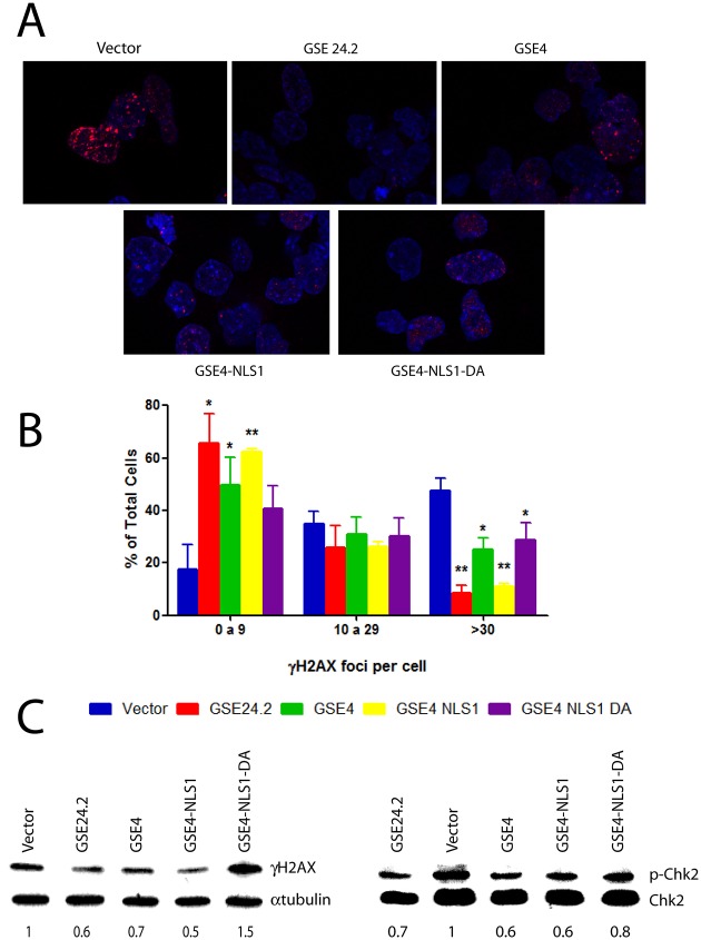
Fig 5. DNA damage protection by the expression of GSE4 and derived peptides.
Panel A, F9-A353V cells were transfected with the pRRL-CMV-IRES-EGFP vector, either empty (vector), or expressing GSE24.2, GSE4, GSE4-NLS1 or GSE4_NLS1-DA (10 μg DNA/106 cells). After 24 hours of transfection cells were fixed and incubated with anti γH2AX and a red-labeled secondary antibody. Nuclear DNA was counterstained with DAPI (blue). Panel B, the amount of γH2AX-expressing foci shown on panel A was determined. The percentage of cells containing 0 to 9, 10 to 29 or more than 30 foci is indicated. More than 200 cells were analyzed in each cell line. Experiments were repeated 3 times with similar results. Panel C, F9_A353V cells were transfected with the plasmids indicated on panel A. Twenty four hours after transfection cells were lysed and the expression of γH2AX, α-tubulin (loading control)(left panel) phosphorylated Chk2 (p-Chk2) and total Chk2 (Chk2)(right panel) analyzed by western blot. Numbers under each blot indicate the relative ɣH2AX/α-tubulin and p-Chk2/Chk2 expression normalized in relation to cells transfected with the empty vector (statistical significance: * p<0.05, **p<0.01, ***p<0.001).

