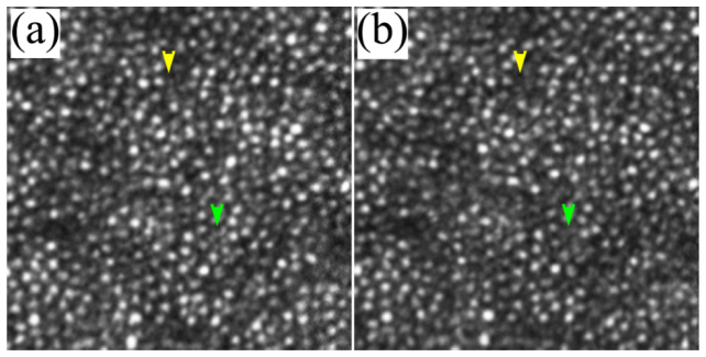
Fig. 11.
AOSLO images taken with 100 Hz AO (a) and 20 Hz AO (b). The images were acquired at 3 degrees eccentricity nasally along the primary horizontal retinal meridian. The size of these images is 256 pixels subtending a field of view of 0.53 degree. Each image is a registered set of 75 AO-corrected frames and has been corrected for distortions due to eye movements. The average center-to-center spacing of cones around the yellow arrowhead is 7.50 μm (measured within a 50 μm X 50 μm area). The average center-to-center spacing of rods is about 3.1 μm (measured from the resolved visible rods). Yellow and green arrowheads indicate improved visibility of rod photoreceptors in the image taken with the high speed 100 Hz AO. DM1 (Hi-Speed DM97-15, ALPAO SAS, France) was used to correct the wave aberration during the image acquisition.

