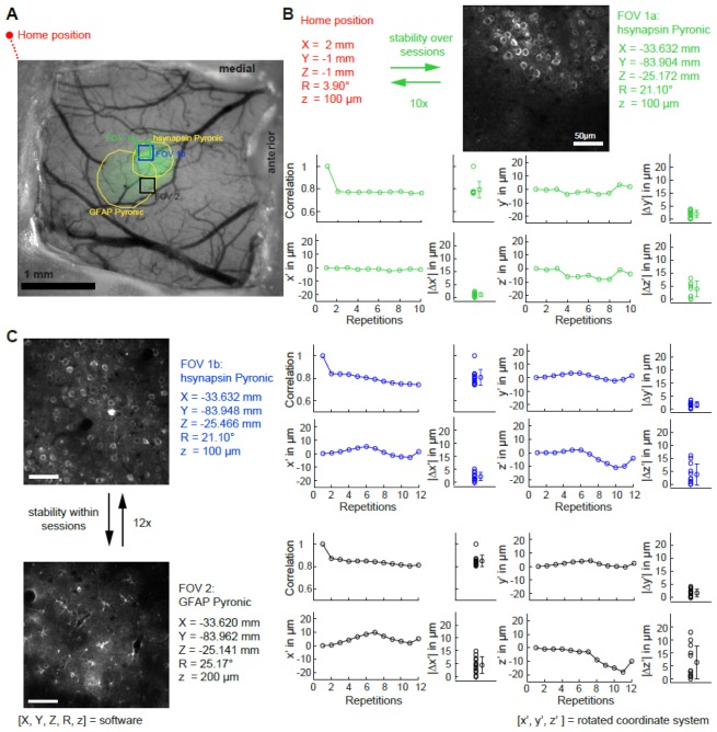
Fig. 3.
Repositioning accuracy in the in vivo situation. (A) Wide field image of chronic window overlaid with indicator expression (green channel; hsynapsin-Pyronic and GFAP-Pyronic). (B) Schematic of the protocol used to measure the accuracy when repositioning the microscope in the same FOV over different sessions (upper panel). Shifts (x’, y’ and z’) and absolute differences (|Δx’|, |Δy’| and |Δz’|) between the initial FOV and subsequent repositioning in the same FOV. (C) Schematic of the protocol used to measure the repositioning accuracy between two FOVs within the same session (left panel). Differences between the initial FOVs and subsequent repositioning in the same FOV (nomenclature same as in (B)).

