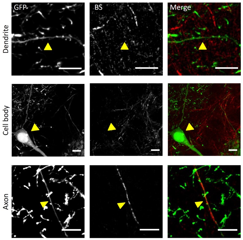
Fig. 2.
Ex vivo BS NIR and two-photon fluorescence imaging. Ex vivo maximum intensity projection (MIP) of stacks (depth = 10 µm) showing the fluorescence and BS NIR signals in a Thy1-GFPM mouse hippocampus. In the upper panel there is a non-reflective GFP-labeled dendrite (arrowhead). Cell bodies (in the middle panel) are also non-reflective. Axons (lower panel) can be both fluorescent and reflective. From left to right: GFP fluorescence, reflectance, merge with GFP in green and reflectance in red. Scale bar, 10 μm.

