Fig. 4.
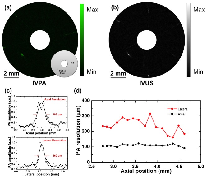
IVPA imaging of 30 μm carbon fiber phantom. (a) IVPA image with schematic of carbon fiber phantom. Black dot indicates 30 μm carbon fiber. (b) IVUS image. (c) The axial and lateral resolutions of IVPA imaging at axial position of 3 mm. (d) The spatial resolutions of IVPA imaging as a function of the axial positions.

