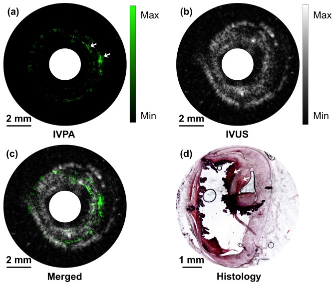
Fig. 6.
High-speed IVPA/IVUS imaging of an excised atherosclerotic human femoral artery. (a) IVPA image. (b) IVUS image. (c) Merged IVPA/IVUS image. (d) Cross-sectional view of the artery histologically assessed for lipids with Oil-Red-O staining. The red indicates the lipid deposition in the artery and the black indicates areas of calcification.

