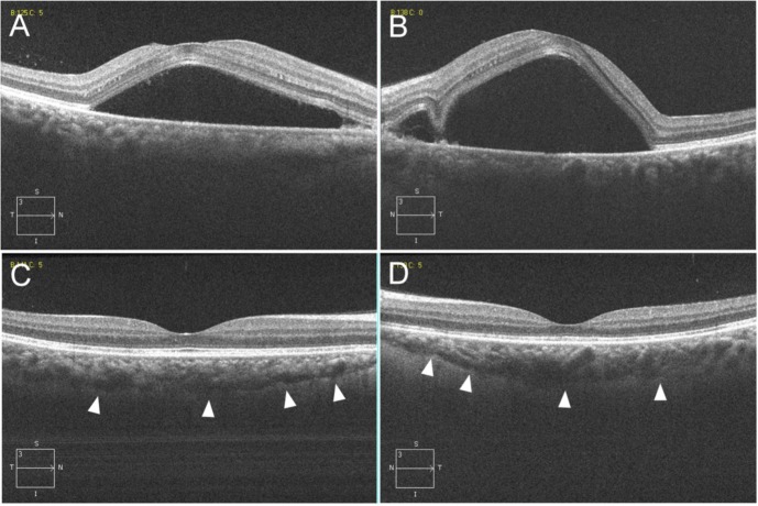
Figure 2.
Horizontal scan images (6.0 mm) from the spectral-domain OCT of the right (A and C) and left eyes (B and D).
Notes: At the initial visit, OCT through the fovea shows a large SRD that included areas of the macula and increased choroidal thickness (which can be observed because of the invisible choroid–scleral interface) in both eyes (A and B). At 40 days after the initial visit, OCT shows not only the disappearance of the SRD, but also marked improvement of the choroidal thickening (which can be observed because of the visible choroid–scleral interface depicted by the arrowheads) (C and D).
Abbreviations: OCT, optical coherence tomography; SRD, serous retinal detachment.

