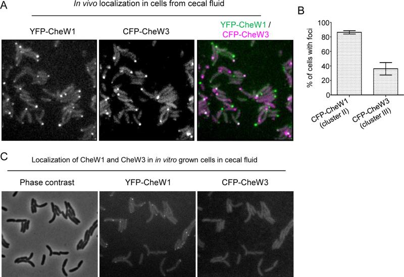
Figure 5. Intracellular localization of YFP-CheW1 and CFP-CheW3 in cells from cecal fluid during V. cholerae colonization of infant rabbits.
(A) Fluorescent micrographs and (B) percentage of cells from cecal fluid with YFP-CheW1 and CFP-CheW3 clusters from cecal fluid infant rabbits infected with V. cholerae strain SR15 (YFP-CheW1, CFP-CheW3). (C) Localization of YFP-CheW1 and CFP-CheW3 in SR15 cells initially grown to exponential phase in LB and then transferred to filtered cecal fluid an imaged after 2hr incubation at 37° with shaking.

