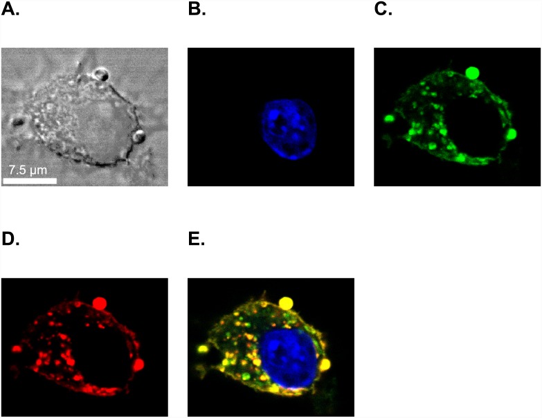
Fig 2. Confocal microscopy confirms the targeted delivery of both lipid and peptide components of GBCA-HDL to the cytoplasm of J774A.1 macrophages.
J774A.1 cells were incubated for 2 h at 37°C with medium only or with medium containing 2.0 μM (as calculated for Rhodamine B) paramagnetic and Rhodamine B-labeled discoidal HDL (dHDL) or spherical HDL (sHDL) synthesized using a 1:1 mixture of oxidized synthetic apo A-I peptides H4 and H6. A small portion of the peptide H4 in the dHDL and sHDL preparations was fluorescently labeled with Dylight 488. (A) Differential interference contrast (DIC) image of a single representative J774A.1 macrophage incubated with sHDL (similar images were obtained for dHDL). The cell was stained with 4’,6-diamino-2-phenylindole (DAPI) dye (blue, B), visualized for Dylight 488-labeled peptide H4 (green, C) and Rhodamine B-labeled lipid (red, D). (E) The merged image shows colocalization of Dylight 488-labeled peptide H4 with Rhodamine B-labeled lipid in the cytoplasm of J774A.1 macrophages, indicating specific uptake of intact sHDL particles into the cytoplasm. White scale bar = 7.5 μM.

