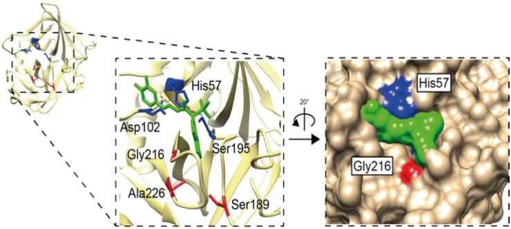
Fig 11. Human chymase structure with inhibitor highlighting the catalytic triad and S1 pocket.
The PDB structure 3N7O was used to visualize specificity-conferring triplet (S1 pocket) of residues 189, 216 and 226, chymotrypsinogen numbering, highlighted in red. The catalytic triad residues His57, Asp102 and Ser195 and shown in blue. The bound inhibitor is highlighted in green in both ribbon and space-filling models to visualize how it sits into the S1 pocket. UCSF Chimera program was used to construct the image with further annotation in Adobe Illustrator (CS6).

