
Figure 5.
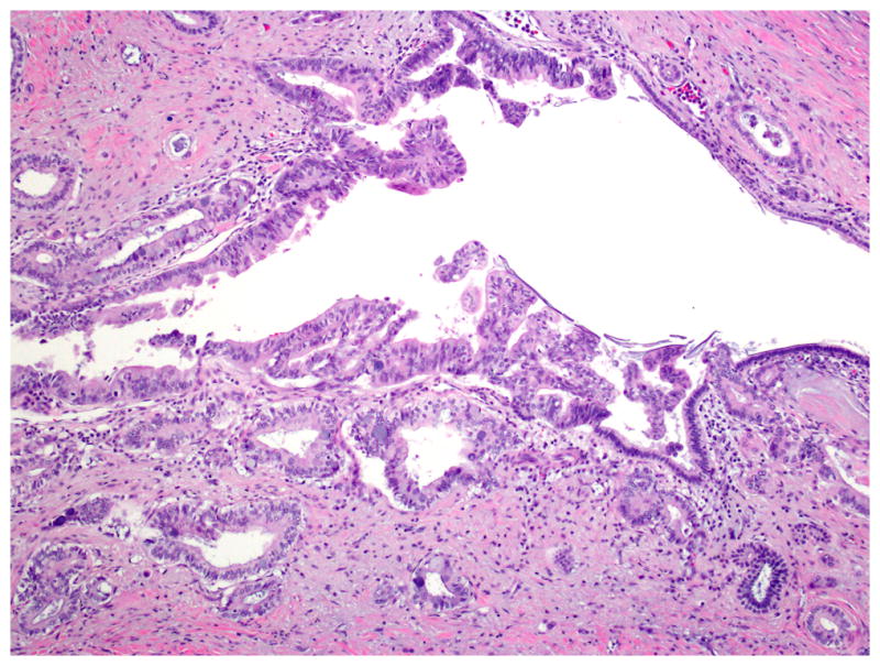
Intraductal spread of invasive carcinoma. Highly atypical epithelia undistinguishable from invasive carcinoma line a duct. Front-formation between normal cuboidal epithelia and the atypical epithelia can be recognized. Hematoxylin and eosin staining. An original magnification was 10×.