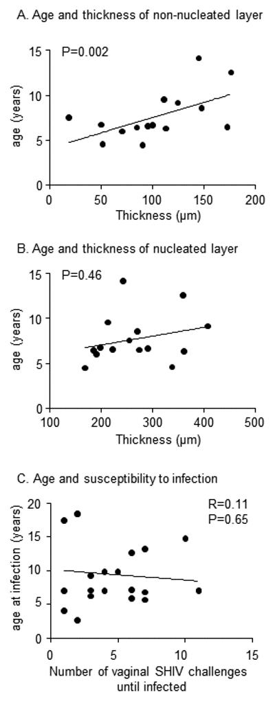
Fig. 5.

Age, vaginal epithelial thickness, and susceptibility to infection. A, B. Scatterplots show the distribution of mean vaginal layer thickness and age of 16 macaques, ranging from 4.5 to 14.2 years. Thickness was evaluated between days 5-20, i.e., when thickness was high, and not thinned by hormonal influences. The line represents the linear relationship; p-value tests the hypothesis of non-zero slope in a multivariable model that controlled for changing levels of thickness over the course of the menstrual cycle C. Scatterplot shows the age of 19 macaques when they became vaginally infected with repeated, low doses of SHIVSF162P3 as described in 24.
