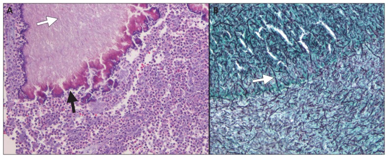
Figure 3:
Histopathologic examination of biopsy specimens from the wedge resection of the left lower lobe of the lung. (A) Hematoxylin and eosin stain showing fungal elements (white arrow) within a bronchiole (black arrow) surrounded by inflammatory cells. (B) Gomori methenamine silver stain showing diffuse fungal elements (white arrow) within a bronchiole (original magnification × 40).

