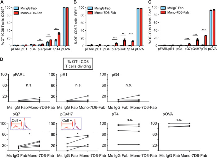
Fig. 3. Mono-7D6-Fab increases T cell responses to weak antigens in vitro.

(A to E) OT-I cells were incubated with the indicated peptides in the presence of Ms IgG Fab or Mono-7D6-Fab for 24 hours (A to C) or 96 hours (D and E). For flow cytometry analysis, OT-I T cells were gated as Thy1.2+Vα2+CD8+. Representative experiments (expressed as percentage) are shown (mean ± SD from triplicate samples): (A) CD69 up-regulation assay (n ≥ 7), (B) IRF4 up-regulation assay (n = 4), (C) T-bet up-regulation assay (n = 2), and (D) CFSE cell division assay. For each peptide stimulation, graphs show paired Ms IgG Fab and Mono-7D6-Fab mean percentages from triplicate samples of dividing OT-I CD8 T cells found in each independent experiment (n = 4). Insets for pQ7 and pQ4H7 depict overlays of the CFSE profiles of Ms IgG Fab–treated (blue line) and Mono-7D6-Fab–treated (red line) samples from one representative experiment. (A to D) *P < 0.05, **P < 0.005, ***P < 0.0005, two-tailed unpaired Student’s t test.
