
Fig. 1. Positive correlation between the expression of CARM1 and MED12 in breast cancer cell lines and human breast tumors.
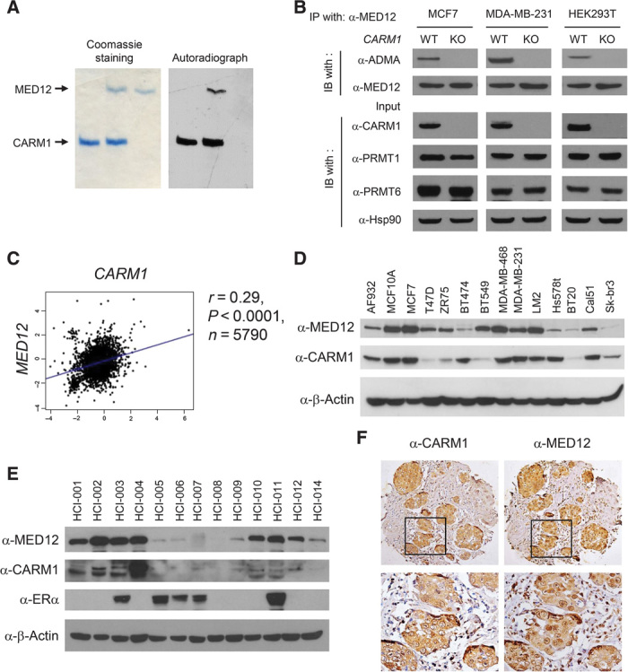
(A) Coomassie brilliant blue staining (left panel) and autoradiograph (right panel) of in vitro methylated MED12 by CARM1 in the presence of [3H]SAM. 3xFLAG-tagged MED12 protein was purified from HEK293T CARM1KO cells. (B) Western blot analyses of immunoprecipitated (IP) MED12 and input lysates from CARM1WT or CARM1KO cell lines using indicated antibodies. WT, wild type; IB, immunoblotting. (C) The Pearson correlation plot depicts the positive correlation between CARM1 and MED12 mRNA expression in 5790 human breast tumor cases collected in the bc-GenExMiner database. (D) Western blot analyses of MED12 and CARM1 proteins in human breast cancer and normal epithelial cell lines. β-Actin was used as an internal control. (E) Western blot analyses of MED12, CARM1, and ERα in patient-derived human breast tumor grafts (36). β-Actin was used as an internal control. (F) Immunohistochemical (IHC) staining of MED12 and CARM1 in human breast tumors.