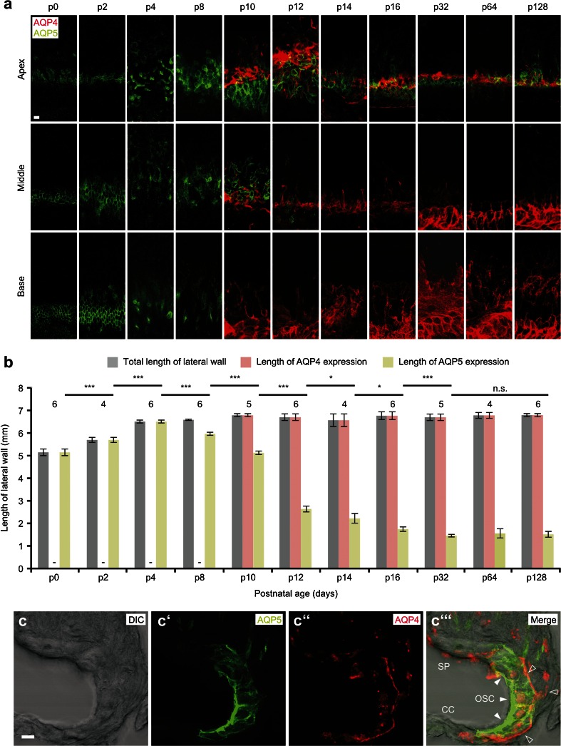
Fig. 1.
Expression of AQP4 and AQP5 in the outer sulcus cell (OSC) region of the mouse cochlear duct during postnatal development. a Immunofluorescence double labeling of AQP4 and AQP5 in spiral ligament whole-mount preparations from the mouse cochlea between postnatal days (p) 0 and p128. Representative confocal images of fluorescence signals were obtained for specimens from the apical (first row), middle (second row), and basal turn (third row) of the cochlea. b Measurements of the total baso-apical length of the spiral ligament (gray bars) and the baso-apical lengths of AQP4 (red bars), and AQP5 fluorescence (green bars) in the outer sulcus region. Length measurements were obtained on whole-mount specimens derived from 4 to 6 cochleae per developmental age (numbers above bars). (Error bars indicate SD, Student’s t test: *p ≤ 0.05; **p ≤ 0.01; ***p ≤ 0.001; n.s. not significant). c–c”’ Confocal images of the subcellular localization of AQP5 (c’, white arrowheads in c”’) in the apical membranes and the cytoplasm; AQP4 (c”, hollow arrowheads in c”’) in the basolateral membranes of OSCs in the apical turn of the mouse (p14) cochlea (SP spiral prominence, CC Claudius cells). Scale bars: (a), 20 μm; (c–c”’), 10 μm

