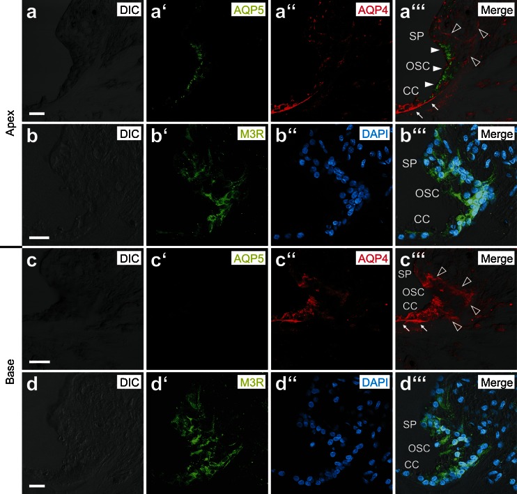
Fig. 5.
Immunolocalization of AQP4, AQP5, and M3R in outer sulcus cells (OSCs) of the adult human cochlea. a–b”’ In the apical turn, AQP4 labeling was localized in the basolateral membranes (hollow arrowheads, a”’), and AQP5 labeling was detected in the apical membranes and the subapical cytoplasm (white arrowheads, a”’) of OSCs. M3R labeling was also detected in OSCs in the apical cochlear turn (b’–b”’). c–d”’ In the basal turn, OSCs exhibited AQP4 labeling in their basolateral membranes (hollow arrowheads, c”’) but were devoid of AQP5 labeling. OSCs in the basal cochlear turn were also immunoreactive for M3R (d–d”’) (White arrows in (a”’ and c”’) indicate AQP4 labeling in the basal membranes of Claudius cells (CCs) in the apical (A”’) and basal turn (C”’)). (SP spiral prominence). Scale bars: 20 μm

