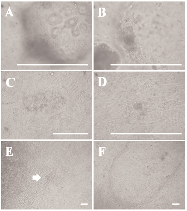
Figure 1.

Outgrowths derived from porcine blastocysts. (A) Morphology of trophectoderm (TE)-like giant cells in a blastocyst surface section. (B) Morphology of an inner cell mass (ICM)-like mass of cells inside a blastocyst. (C) Giant cells attached to feeder layer cells 3 days after blastocyst seeding. (D) The mass of cells presumed to be the ICM is attached to the feeder layer. (E) Morphology of an embryonic stem (ES)-like primary colony derived from a porcine embryo. (F) Late morphology of an ES-like primary colony derived from a porcine embryo. Scale bars = 100 μm.
