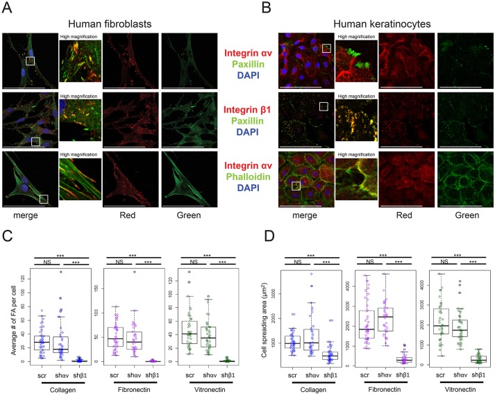
Fig. 2.
Integrin αv does not localize to focal adhesions in keratinocytes. (A,B) Representative images of human fibroblasts (A) or human keratinocytes (B) stained for integrin ɑv, integrin β1 and paxillin, and/or incubated with phalloidin, then imaged with confocal microscopy. (C) Box plots showing quantification of the number of focal adhesions per cell for keratinocytes infected with a scramble hairpin, an integrin αv hairpin (shαv) or an integrin β1 hairpin (shβ1), and seeded onto coverslips coated with collagen, fibronectin or vitronectin (n=30–40 cells per condition, box-plot whisker ends are at the 1.5 interquartile range, boxes are at the 1st and 3rd quartile, and the line is at the median). P=2.45×10⁻¹² (collagen), 8.15×10⁻¹⁵ (fibronectin), 2×10⁻¹⁶ (vitronectin) using one-way ANOVA. (D) Box plots showing quantification of cell spreading area for the same cells shown in C. P=3.79×10⁻⁹ (collagen), 2×10⁻¹⁶ (fibronectin), 2×10⁻¹⁶ (vitronectin) using one-way ANOVA. Scale bars: 100 μm. ***P<0.0005; NS, not significant.

