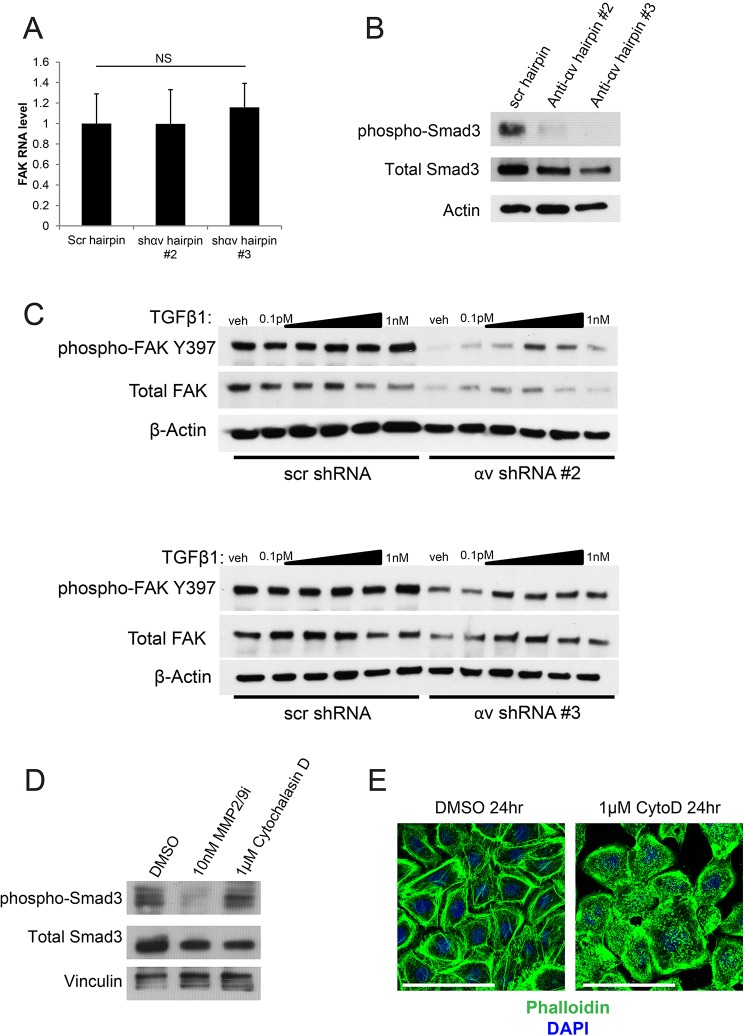
Fig. 4.
TGFβ signaling is partially responsible for the regulation of FAK signaling by integrin αv. (A) qPCR analysis of FAK transcript levels in keratinocytes that had been infected with the indicated shRNAs. NS, not statistically significant, measured with one-way ANOVA (P=0.906). Scr, scrambled shRNA; shɑv #2 and #3, two independent shRNAs against integrin ɑv. (B) Western blot showing signaling pathway changes in keratinocytes that had been infected with the indicated shRNAs. anti-ɑv hairpin #2 and #3, two independent shRNAs against integrin ɑv. (C) Western blot showing signaling pathway changes upon addition of varying doses of TGFβ1 in keratinocytes that had been infected with the indicated hairpins. TGFβ1 doses range from 0.1 pM to 1 nM, increasing by tenfold each time. Veh, vehicle. (D) Western blot showing signaling pathway changes in keratinocytes upon treatment with 10 nM of a MMP2 and MMP9 inhibitor (MMP2/9i), or 1 µM cytochalasin D treatment for 24 h. (E) Phalloidin staining of keratinocytes treated with control (DMSO) or 1 µM cytochalasin D (CytoD) for 24 h. Scale bars: 100 μm.

