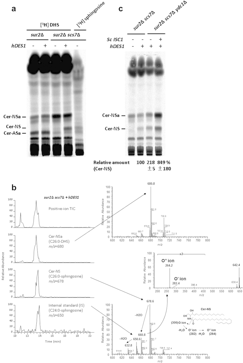
Figure 3. Ceramide-NS is formed in the sur2Δ scs7Δ mutant expressing human DES1.
(a,c) Cells were labeled with [3H]DHS overnight at 25 °C. The labeled lipids were extracted, subjected to mild alkaline hydrolysis, spotted on borate-impregnated TLC plates, and developed with solvent system III. Radioactivity of ceramide-NS in (c) was quantified and expressed as the relative amount to that in sur2Δ scs7Δ mutant expressing human DES1.Data represent the average of two independent experiments with error bars denoting the range of the two experiments. (b) HPLC/ion-trap mass spectroscopy of ceramides in the sur2Δ scs7Δ mutant expressing human DES1. (Left) Total ion current (TIC) and ion chromatograms of the most intense signals of Cer-NSa (C26:0), Cer-NS (C26:0), and internal standard Cer-NS (C24:0) in the positive-ion mode. (Right) Mass spectra of Cer-NSa (upper panel) and Cer-NS (lower panel). The lower inset shows the fragmentation patterns of Cer-NS at the amide linkage12. A MS-MS spectrum of the m/z 660.8 ions (middle panel).

