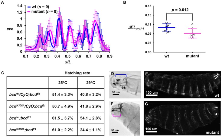
Fig. 6.
Developmental defects exhibited by mutant embryos. (A) Normalized eve intensity profile from wt and mutant embryos. (B) ΔELeve3-4 value in individual wt and mutant embryos. (C) The Bcd mutant reduces the hatching rate in a gene dosage- and temperature-dependent manner (29°C versus 25°C for two versus one copy of the mutant transgene). (D,E) The cuticle pattern of an embryo from the bcdwt;bcdE1 mother at 29°C showing (D) the head structure from Nomarski microscopy and (E) the denticle pattern of the whole body under dark-field microscopy. (F,G) Cuticle patterns of an embryo from the bcdK308A;bcdE1 mother at 29°C. Note a shortening in mediate tooth (mt) and epistomal sclerite (es) and a denticle fusion in this embryo. Brackets indicate the spans from mt to the end of es.

