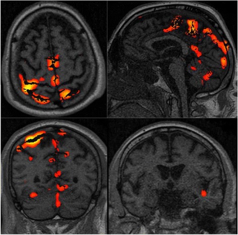
Fig. 4.

Functional magnetic resonance imaging (fMRI) (21 days after cardiac arrest (CA)) of patient 12 of our series who had a good outcome, showing the brain activation of the somatosensory, motor, premotor, left insula and cerebellum areas. Some artifacts are also visible. The neurophysiological recording performed at 24 h after CA showed pain-related somatosensory evoked potentials (ML-SSEP) and a diffuse, slowing, nonreactive electroencephalogram (EEG). The subsequent EEG patterns were characterized by epilectiform activity. This patient regained consciousness at 33 days after CA
