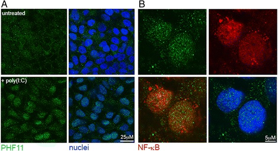
Fig. 3.

Nuclear localization of PHF11 is induced by poly(I:C). a Confocal microscopy of PHF11 localization in HaCaT keratinocytes on day 4 of culture in the absence of poly(I:C) (untreated), or after a 24-h poly(I:C) treatment (+poly(I:C)). b Higher magnification image of the nuclei of cells treated with poly(I:C) and stained for PHF11 (green), NF-κB (red). A merged image of PHF11 and NF-κB (lower left), as well as DAPI-stained nuclei (lower right) is shown
