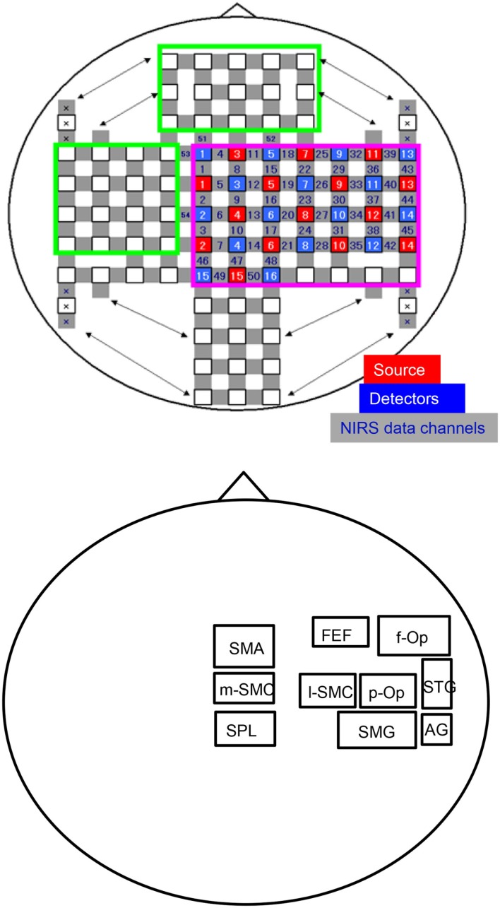
Figure 1.
An illustration of the arrangement of the optodes (sources and detectors) and recording channels (A) and a schema of the 10 regions of interest (ROIs) for the group-averaged NIRS data analysis (B). f-Op, right frontal operculum/inferior frontal gyrus; p-Op, right parietal operculum; FEF, frontal eye field; SMG, right supramarginal gyrus; AG, right angular gyrus; STG, right superior temporal gyrus; l-SMC, lateral part of the sensorimotor cortex in the right hemisphere; m-SMC, medial part of the sensorimotor cortex; SPL, superior parietal lobule; SAC, somatosensory association cortex; SMA, supplementary motor area.

