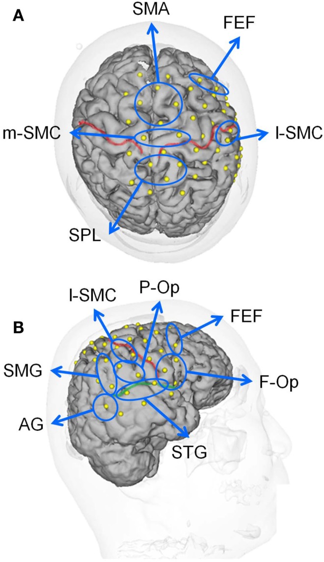
Figure 2.
Locations of the recording channels and 10 regions of interest (ROIs) on a 3-D MRI reconstruction of the brain of one of the subjects. (A,B) indicate the top and right views of the brain, respectively. The red and green lines indicate the central sulcus and right Sylvian fissure, respectively. f-Op, right frontal operculum/inferior frontal gyrus; p-Op, right parietal operculum; FEF, frontal eye field; SMG, right supramarginal gyrus; AG, right angular gyrus; STG, right superior temporal gyrus; l-SMC, lateral part of the sensorimotor cortex in the right hemisphere; m-SMC, medial part of the sensorimotor cortex; SPL, superior parietal lobule; SAC, somatosensory association cortex; SMA, supplementary motor area.

