FIGURE 5.
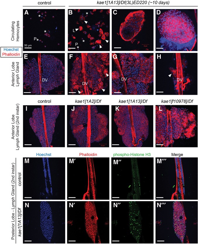
Hematopoietic overgrowth and blood cell defects in kae1 mutants. All samples were stained with Hoechst (blue) to label DNA and rhodamine phalloidin (red) to label F-actin; all scale bars, 50 µm. Control animals were TM6B siblings of crosses used to generate kae1/Df(3L)ED220 larvae and are thus heterozygous for kae1. (A) Control hemolymph shows the typical profile in which most circulating hemocytes are plasmatocytes ([P] asterisks). (B) kae1[1A13] hemizygous hemolymph shows increased density of blood cells, as well as numerous lamellocytes, often present in clusters ([L] arrowheads). Lamellocytes (∼40–50 µm in diameter) are larger than plasmatocytes (10 µm in diameter). (C) Large aggregates bearing organized structure were observed in circulation as were overt tumors that are packed with plasmatocytes and lamellocytes (D). The latter correspond to the melanotic masses observed in live “black spot” larvae. (E–H) Anterior lobe of lymph gland from third instar in control (E) and from aged kae1/Df(3L)ED220 larvae (F–H); the dorsal vessel (DV) is labeled. The kae1 mutants exhibit strong overcommitment to lamellocyte fate in the lymph gland (F) and typically have overgrown lobes (G). Frequently, the anterior lobe was observed in the process of dispersal or was detached entirely from the dorsal vessel (H). (I–L) Analysis of anterior lymph gland lobes in second instar larvae. Compared with control (I), all kae1 mutants exhibit ectopic lamellocytes (J–L). The hypomorphs [1A2] (J) and [1A13] (K) are relatively normally sized, but the null [f01978] (L) is strongly overgrown. (M,N) Analysis of mitotic cells (labeled with anti-phospho-Histone H3, green) in posterior lobes of lymph glands from control (M) and [1A13]/Df (N). The kae1 mutant has overgrown posterior lobes that harbor more actively dividing cells.

