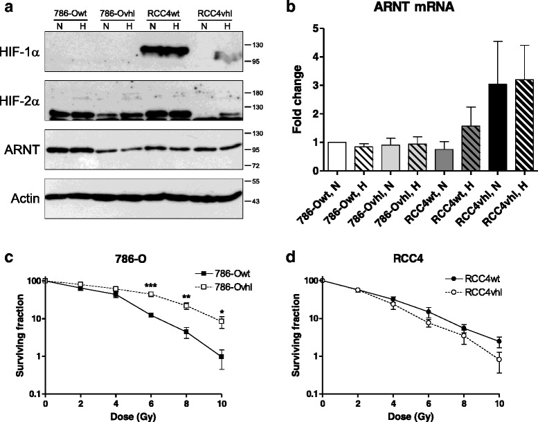
Fig. 2.

ARNT expression in renal carcinoma cells and response to radiation. a 786-O and RCC4 wildtype (wt) and pVHL expressing (vhl) cells respectively were seeded on Petri-dishes followed by exposure to normoxia (N) or hypoxia (H, 3 % O2) for 8 h. Protein levels of HIF-1α, HIF-2α and ARNT were assayed by Western Blotting. Actin levels were determined for loading control. Protein masses are indicated on the right in kDa. Representative result of n = 3 independent experiments. b ARNT mRNA expression in 786-Owt, 786-Ovhl, RCC4wt and RCC4vhl cells exposed to normoxia (N) or hypoxia (H, 3 % O2) for 8 h. ARNT- and B2M (endogenous control) mRNA were measured using TaqMan® chemistry and normalized to normoxic 786-Owt cells according to the ∆∆Ct method. Fold changes of ARNT mRNA levels are represented as mean +/− SEM of n = 3 independent experiments. c Clonogenic survival assays of irradiated 786-Owt and 786-Ovhl cells. n = 6, mean +/− SEM, unpaired t-test; (d) Clonogenic survival assays of irradiated RCC4wt and RCC4vhl cells. n = 5–6, mean +/− SEM, unpaired t-test
