Fig. 3.

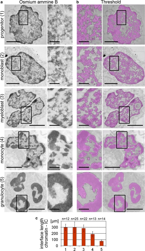
Chromatin landscapes of nuclei of various myelopoietic differentiation stages visualized by TEM in osmium ammine B stained physical sections and assessment of the chromatin/IC interface length. a A transition from a fine network of CDCs/IC channels toward a dense and more lump-like pattern was observed with progressive myeloid differentiation. b Thresholded masks for the delineation of osmium ammine B stained chromatin (pink) and the IC (gray) in the respective sections. Scale bars 2 µm, insets 0.5 µm. c The interface length between the thresholded chromatin and the IC is reduced in differentiated cell types (1 progenitor; 2 monoblast; 3 myeloblast; 4 monocyte; 5 granulocyte). n Number of analyzed nuclei; error bars standard deviation; p < 0.001 for monocytes and granulocytes versus respective precursors and progenitors
